Why the eye is better than a camera
The human eye long ago solved a problem common to both digital and film cameras: how to get good contrast in an image while also capturing faint detail.

Nearly 50 years ago, physiologists described the retina’s tricks for improving contrast and sharpening edges, but new experiments by University of California, Berkeley, neurobiologists show how the eye achieves this without sacrificing shadow detail.
“One of the big success stories, and the first example of information processing by the nervous system, was the discovery that the nerve cells in the eye inhibit their neighbors, which allows the eye to accentuate edges,” said Richard Kramer, UC Berkeley professor of molecular and cell biology. “This is great if you only care about edges. But we also want to know about the insides of objects, especially in dim light.”
Kramer and former graduate student Skyler L. Jackman, now a post-doctoral fellow at Harvard University, discovered that while light-sensitive nerve cells in the retina inhibit dozens of their close neighbors, they also boost the response of the nearest one or two nerve cells.
That extra boost preserves the information in individual light detecting cells – the rods and cones – thereby retaining faint detail while accentuating edges, Kramer said. The rods and cones thus get both positive and negative feedback from their neighbors.
“By locally offsetting negative feedback, positive feedback boosts the photoreceptor signal while preserving contrast enhancement,” he said.
Jackman, Kramer and their colleagues at the University of Nebraska Medical Center in Omaha report their findings today (Tuesday, May 3) in the journal PLoS Biology. Kramer also will report the findings today at the 2011 annual meeting of the Association for Research in Vision and Ophthalmology in Ft. Lauderdale, Fla.
From horseshoe crabs to humans
The fact that retinal cells inhibit their neighbors, an activity known as “lateral inhibition,” was first observed in horseshoe crabs by physiologist H. Keffer Hartline. That discovery earned him a share of the 1967 Nobel Prize in Physiology or Medicine. This form of negative feedback was later shown to take place in the vertebrate eye, including the human eye, and has since been found in many sensory systems as a way, for example, to sharpen the discrimination of pitch or touch.
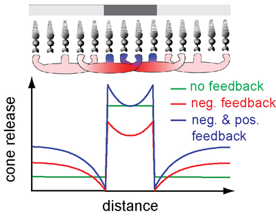
Lateral inhibition fails, however, to account for the eye’s ability to detect faint detail near edges, including the fact that we can see small, faint spots that ought to be invisible if their detection is inhibited by encircling retinal cells.
Kramer noted that the details of lateral inhibition are still a mystery half a century after Hartline’s discovery. Neurobiologists still debate whether the negative feedback involves an electrical signal, a chemical neurotransmitter, or protons that change the acidity around the cell.
“The field is at an impasse,” Kramer said. “And we were surprised to find this fundamental new phenomenon, despite the fact that the anatomy of the retina has been known for more than 40 years.”
The retina in vertebrates is lined with a sheet of photoreceptor cells: the cones for day vision and the rods for night vision. The lens of the eye focuses images onto this sheet, and like the pixels in a digital camera, each photoreceptor generates an electrical response proportional to the intensity of the light falling on it. The signal releases a chemical neurotransmitter (glutamate) that affects neurons downstream, ultimately reaching the brain.
Unlike the pixels of a digital camera, however, photoreceptors affect the photoreceptors around them through so-called horizontal cells, which underlie and touch as many as 100 individual photoreceptors. The horizontal cells integrate signals from all these photoreceptors and provide broad inhibitory feedback. This feedback is thought to underlie lateral inhibition, a process that sharpens our perception of contrast and color, Kramer said.
The new study shows that the horizontal cells also send positive feedback to the photoreceptors that have detected light, and perhaps to one or two neighboring photoreceptors.
“Positive feedback is local, whereas negative feedback extends laterally, enhancing contrast between center and surround,” Kramer said.
Electrical vs. chemical signals
The two types of feedback work by different mechanisms, the researchers found. The horizontal cells undergo an electrical change when they receive neurotransmitter signals from the photoreceptors, and this voltage change quickly propagates throughout the cell, affecting dozens of nearby photoreceptors to lower their release of the glutamate neurotransmitter.
The positive feedback, however, involves chemical signaling. When a horizontal cell receives glutamate from a photoreceptor, calcium ions flow into the horizontal cell. These ions trigger the horizontal cell to “talk back” to the photoreceptor, Kramer said. Because calcium doesn’t spread very far within the horizontal cell, the positive feedback signal stays local, affecting only one or two nearby photoreceptors.
The discovery of a new and unsuspected feedback mechanism in a very well-studied organ is probably related to how the eye is studied, Kramer said. Electrodes are typically stuck into the retina to both change the voltage in cells and record changes in voltage. Because the new signal is chemical, not electrical, it would have been easily missed.
Jackman and Kramer found the same positive feedback in the cones of a zebrafish, lizard, salamander, anole (whose retina contains only cones) and rabbit, proving that “this is not just some weird thing that happens in lizards; it seems to be true across all vertebrates and presumably humans,” Kramer said.
The research was supported by the National Institutes of Health and the organization Research to Prevent Blindness.
Coauthors with Kramer and Jackman are Norbert Babai and Wallace B. Thoreson of the Department of Ophthalmology at the University of Nebraska Medical Center and James J. Chambers of the Department of Chemistry at the University of Massachusetts, Amherst.
For more information:
- PLoS Biology article, “A Positive Feedback Synapse from Retinal Horizontal Cells to Cone Photoreceptors”
- Richard Kramer’s laboratory web site
- Photoswitches could restore sight to blind retinas (2006 press release)