Unusual coronavirus protein is potential drug target to fight COVID-19

The SARS-CoV-2 virus contains a gene that codes for a strange protein that could be a good target for drugs to fight COVID-19 and possibly other coronavirus infections, according to a new study from the University of California, Berkeley.
When the virus injects its genome into a cell, the so-called ORF3a gene is expressed to make a protein that moves to the surface of the cell and looks like an ion channel — a passage all cells have for ions like calcium to move in and out as they communicate with other cells.
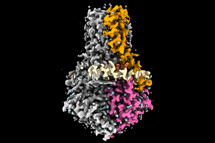
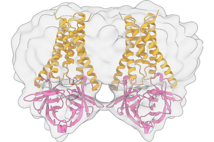
But UC Berkeley researchers, who began investigating the protein’s structure with cryogenic electron microscopy (cryoEM) in January of last year, immediately after the pandemic began, discovered that the protein is nothing like other ion channels known to science. For one thing, it’s half a channel — it only pierces the cell membrane halfway. It also has an unusual fold not seen in other ion channels.
While no one knows why the virus carries a gene to make an ion channel — and a strange one, at that — researchers have found that by knocking out the ORF3a gene in SARS-CoV-2 and a related ORF3a gene in the original SARS virus, SARS-CoV-1, the severity of the disease is reduced, at least in animal models. This, the researchers say, makes it a good target for drugs to reduce the severity of human coronavirus infections. A high-resolution cryoEM structure published this week by the UC Berkeley team provides key information needed to find such drugs.

“It wasn’t clear how conserved this protein was among coronaviruses, but once we solved the structure, we could look to see if other genes in other coronaviruses are likely to adopt the same fold,” said Stephen Brohawn, one of the senior authors of the study and a UC Berkeley assistant professor of molecular and cell biology. “And it turns out that in all of the coronaviruses that circulate among bats and can infect humans, these 3a proteins exist. So, it could be an even broader possible target than we had thought at the beginning of the project.”
Brohawn and his UC Berkeley colleagues have already identified a drug that blocks the ion channel — though it also blocks other ion channels — and have identified mutations in the ORF3a gene that alter channel function.
“This shows, in principle, that one can find molecules that block this activity,” Brohawn said. “Now one of the goals that we have is to screen for small molecules that block the channel and that are specific for blocking 3a compared to other ion channels.”
High-resolution cryoEM structures
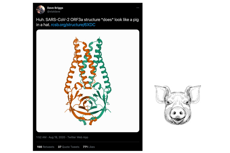
The team’s preliminary results were posted on the open access preprint server bioRxiv in June of 2020, and the final results — with a much higher resolution cryoEM structure for the 3a protein — were published yesterday in the journal Nature Structural & Molecular Biology. The original online paper drew attention from those using computational approaches to find drugs to fight COVID-19, but it also helped improve a computer algorithm — Google DeepMind’s AlphaFold — that predicts a protein’s 3D structure from its amino acid sequence.

Brohawn, who is member of UC Berkeley’s Helen Wills Neuroscience Institute, uses cryoEM to study the structures of ion channels in neurons. For this project, he teamed up with the lab of Diana Bautista, UC Berkeley professor of molecular and cell biology, who uses other methods to study ion channels, specifically those involved in chronic itch, pain and inflammatory disease. Together, the two labs provided three different pieces of evidence that the 3a protein does act as an ion channel. Though they demonstrated this in artificially-made lipid membranes called liposomes, these membranes are chemically similar to the membranes of cells, so presumably the proteins also work as ion channels in coronavirus-infected cells.
The odd structure of the 3a protein — specifically, its blind pore, which is totally unlike typical ion channels that contain a tunnel that goes completely through the membrane to allow ions easy passage in and out — suggests that ions may instead diffuse down grooves along the outside of the protein.
According to Brohawn, the 3a protein is the smallest membrane protein channel that has been imaged by cryoEM, which has rapidly become the best way to determine the 3D structure of molecules, down to the level of individual atoms and the water molecules that surround them. Brohawn continues to research the 3a protein and two other potential ion channels — called E and 8a —in the SARS-CoV-2 genome, both to understand how they work and to find potential drugs to inactivate them.
Bautista continues investigating how the virus uses these ion channels to take over human and animal epithelial cells and neurons. Many viruses interfere with calcium signaling in cells, which may help them take over the cells’ molecular machinery and force them to make millions of copies of the virus instead of daily cellular housekeeping.
“We thought 3a was the most understudied and potentially the most interesting of these proteins and something we could probably make a dent in, if we jumped on it right away,” Brohawn said. “We were in a good position move quickly on this, and it was really exciting to see it come together that fast.”
Co-authors of the study are David Kern, Ben Sorum, Sonali Mali, Christopher Hoel, Savitha Sridharan, Jonathan Remis and Daniel Toso of UC Berkeley and Abhay Kotecha of Thermo Fisher Scientific in the Netherlands. The work was funded in part by a Fast Grants award from Emergent Ventures at the Mercatus Center at George Mason University.
RELATED INFORMATION
- Cryo-EM structure of SARS-CoV-2 ORF3a in lipid nanodiscs (Nature Structural & Molecular Biology)
- Stephen Brohawn’s lab website
- Diana Bautista’s lab website
