New MRI Method Offers Deeper Insight Into Brain Physiology
Technique traces blood flow sources ‘in reverse’ to study brain function and disease

The venous system maintains the health of our brains by removing deoxygenated blood and other waste products, but its complexity and variability have made scientific study difficult. Now, a UC Berkeley-led team of researchers has developed an innovative MRI technique that may expand our understanding of this critical system.
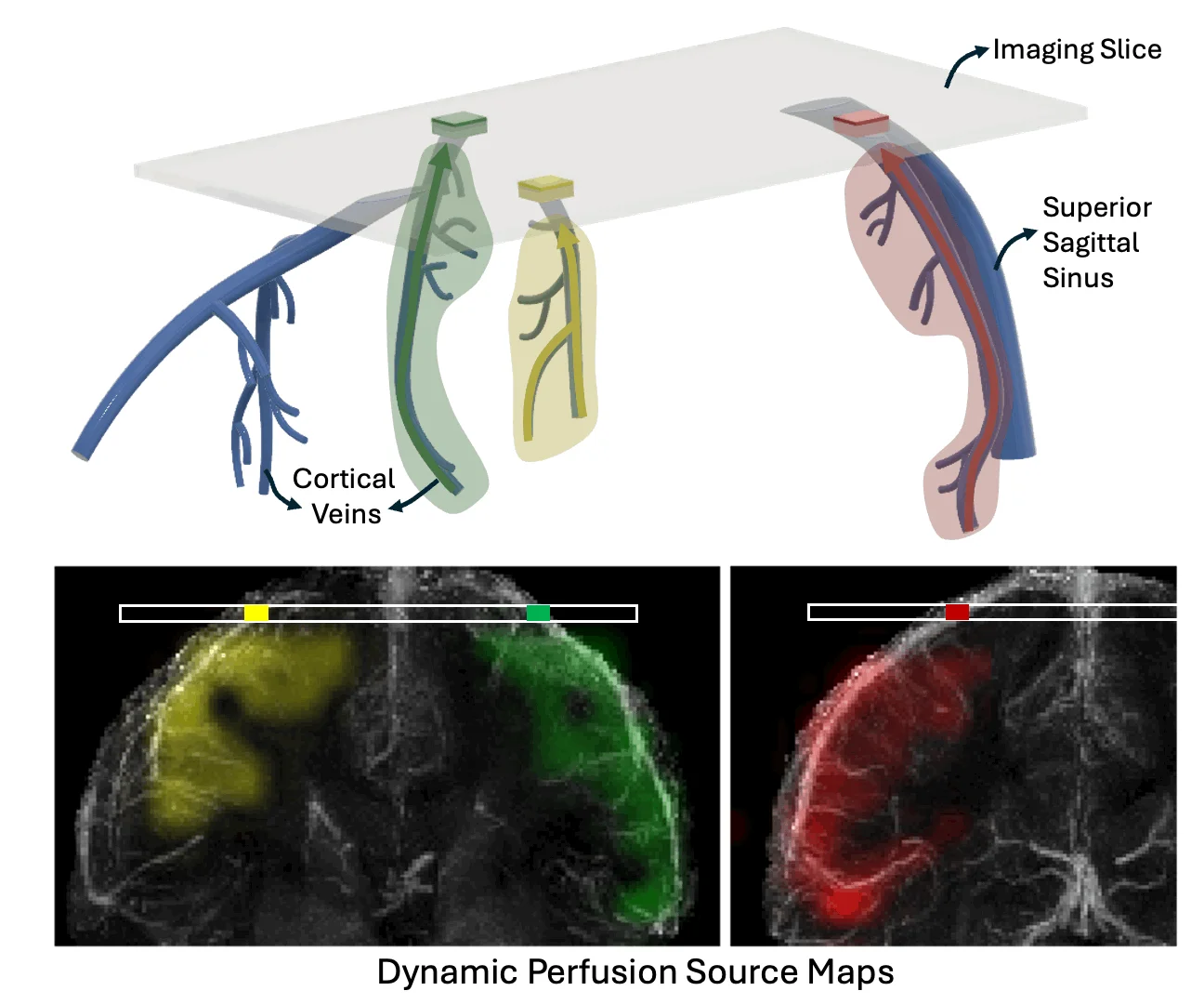
In a study published in Nature Communications, the researchers demonstrate how their new imaging method, Displacement Spectrum (DiSpect) MRI, maps blood flows “in reverse” to reveal the source of blood in the brain’s veins. This approach could help answer long-standing questions about brain physiology as well as provide a safer, more efficient way to diagnose disease.
Like some current MRI methods, DiSpect uses the water in our blood as a tracing agent to map perfusion, or blood flow, in the brain. The water’s hydrogen atoms possess a quantum mechanical property called spin and can be magnetized when exposed to a magnetic field, like an MRI scanner. But what makes DiSpect unique is its ability to track the “memory” of these nuclear spins, allowing it to map blood flow back to its source.
“We can tag information onto spins in the blood, knowing that they store that information for three, four, five seconds, or so,” said Michael Lustig, professor of electrical engineering and computer sciences and the study’s principal investigator. “In that time, the tagged blood will have traveled, draining from the brain’s capillaries and smaller veins into larger veins. And when we image it at the larger veins, we can decode the information to determine where it came from.”

In their study, the researchers demonstrated how the images produced with DiSpect can be used to trace the vein’s blood flow back to the specific region of the brain that it’s draining, known as its territory.
In addition, they showed how their technique can detect changes in blood flow. For example, DiSpect proved sensitive enough to detect global blood flow changes, such as those caused by caffeine intake. It also detected more subtle, localized changes induced by brain activity during basic movements like hand squeezing.
During these simple motor tasks, DiSpect was able to image the redistribution of blood from the passive area of the brain to the region exhibiting neuronal activity.
“This is an occurrence that has been widely hypothesized but difficult to directly observe using conventional MRI methods, like functional MRI [fMRI],” said lead author Ekin Karasan, a postdoctoral researcher in the Department of Electrical Engineering and Computer Sciences. “DiSpect could potentially help us gain a deeper understanding of blood flow redistribution as it relates to brain activity — a phenomenon known as arterial blood stealing.”
As such, DiSpect may prove useful in shedding light on basic neuroscience questions tied to neurovascular coupling, the mechanism by which blood flow changes in response to neuronal activity.
“When it comes to detecting brain activity, our method can help us determine whether the neurons are driving signal changes, or whether they are being driven by local perfusion of blood movement,” said co-author Chunlei Liu, a professor in the Department of Electrical Engineering and Computer Sciences and the Helen Wills Neuroscience Institute. “Unlike other MRI methods, DiSpect allows you to pin down where the local perfusion changes are happening, which could help advance our understanding of these mechanisms.”
DiSpect’s reverse imaging technique may also lead to more effective strategies for treating vascular and neurological conditions. “This approach can help us better understand how changes to the venous structure and venous dynamics may contribute to disease — valuable knowledge that can help guide treatment decisions,” said Liu.
Karasan hopes that DiSpect can someday be used to assess the health risk of arteriovenous malformations. These bundles of abnormally formed blood vessels are like tangled balls of yarn and can occur in the brain and elsewhere. Currently, clinicians must spend hours inserting a catheter and contrast into each tiny artery to identify which one is feeding the malformation — and to determine whether treatment is even necessary.
“DiSpect could provide a no-contrast, non-invasive and less risky detection method,” said Karasan. “Starting at the veins that are draining this malformation, we could use reverse imaging to identify which artery the blood is coming from — [and] then figure out whether intervention is required.”
According to Lustig, this work shows how a technology that operates at the atomic scale has the power to help us answer some big scientific questions.
“You’re interacting with the smallest things in the universe, the nucleus of hydrogen atoms. But through the miracle of MRI, you can infer from these atoms where blood flows in veins, the sources of it, and see brain activity, and modulations of it,” he said. “Every time I think about it, it’s just mind-blowing.”