
Research Bio
Physical, Biophysical and Bioanalytical Chemistry — Richard Mathies has developed a variety of modern laser-based spectroscopic techniques to study excited-state reaction dynamics in photoactive proteins and to develop novel microfabricated chemical and biochemical analysis devices for terrestrial and extra-terrestrial applications.
Ultrafast Vibrational Spectroscopy of Reaction Dynamics. Mathies has used resonance Raman vibrational spectroscopy to study photochemical reaction dynamics of energy and information transducing systems especially the primary photochemistry of rhodopsins. Most recently his group developed the novel technique of Femtosecond Stimulated Raman Spectroscopy or FSRS that enables the acquisition of femtosecond time-resolved vibrational structure snapshots of reacting molecules.
Research Expertise and Interest
genomics, biophysical, bioanalytical, physical chemistry, laser spectroscopy, resonance Raman, excited-state reaction dynamics photoactive proteins, rhodopsins, microfabricated chemical biochemical analysis devices, forensics, infectious disease detection
In the News
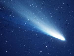
Evidence that comets could have seeded life on Earth
A new experiment simulating conditions in deep space reveals that the complex building blocks of life could have been created on icy interplanetary dust and then carried to Earth, jump-starting life.
$3.5 million gift from Dow to develop sustainable chemistry education
With the support of the gift from The Dow Chemical Company Foundation, the College of Chemistry will rebuild its undergraduate teaching labs and design a new curriculum.
Agilent helps launch new synthetic biology center
Agilent Technologies Inc. has signed up to support the newly launched Synthetic Biology Institute (SBI), which will help advance efforts to engineer cells and biological systems in ways that could transform health and medicine, energy, the environment and new materials.
Teaching
Seminars for Graduate Students [CHEM 298 - 052]
Research for Graduate Students [CHEM 299 - 034]
Seminars for Graduate Students [CHEM 298 - 046]
Research for Graduate Students [CHEM 299 - 045]
Research [BIOPHY 292 - 001]
Seminars for Graduate Students [CHEM 298 - 052]
Research for Graduate Students [CHEM 299 - 034]