UC Berkeley-UCSF Faculty Collaboration Program
About
The UC Berkeley-UCSF Faculty Collaboration Program strives to reward innovative research approaches that take advantage of and promote the convergence of the biomedical, physical and engineering fields encouraging scientists to move into new fields and cross into other areas of convergence in order to realize the full potential for achieving transformative scientific breakthroughs.
To advance this fertile research area, the program facilitates innovation and faculty collaboration between UC Berkeley and UC San Francisco through a prestigious collaboration program. The program currently provides support for one scientist, ideally alternating from each of the two institutions, to collaborate with colleagues at the other institution each year.
In this way, the program ensures that faculty research agendas are enriched by a widened circle of immediate colleagues and collaborators promoting the integration of research communities across different disciplines.
Applications are submitted directly to the Office of the Vice Chancellor for Research via this online application form.
How to Apply
Applications for the 2025 cycle were due February 27, 2025 (6:00 PM).
Awards will be for $50,000 for one year. Please review the proposal guidelines and upload your application materials below. If you have questions about overall program parameters and guidelines, please consult the program's FAQs. If you have additional questions, please contact research@berkeley.edu.
The program is restricted to full-time UC Berkeley and UCSF faculty at the Assistant, Associate, and Full Professor ranks of any professional series (including, but not limited to adjunct, in-residence, health sciences, clinical, and professor of clinical optometry).
Proposals are reviewed by faculty peers from STEM departments from both campuses. Please be cognizant that reviewers will come from different STEM disciplines, so proposals should be accessible for a general scientific community.
Please submit the following as a single pdf via this application form.
- Cover Page & Abstract (one page max)
- Applicant and collaborator’s name
- Rank/Title and affiliations for both collaborators
- Title of proposed research project
- Project abstract, which should clearly outline the overarching goals of the research proposal in a way that is accessible to a lay-audience (200 words max).
- Research Project Description (up to four pages, excluding references, CV, and budget justification)
- It should promote the convergence of biomedical, physical, and engineering fields to foster the innovations necessary for meeting the health care challenges of the future.
- Please be sure to provide details on the innovative nature of the proposed work and how the collaboration across campuses will occur.
- Budget Justification: A detailed budget is not required, however an outline/justification of how the support will enhance the project is required. The funding may be used to support research expenses, including but not limited to:
- performing experiments
- analyzing research data
- supporting workshops
- colloquia
- research travel
- supporting postdocs, graduate, or undergraduate research assistants
- PI salary, including summer salary, is not an allowable expense.
- You do not need to deduct any overhead
- The application does not need to be reviewed by the Sponsored Projects Office (UCB) or Contracts & Grants (UCSF)
- CV or Biosketch (abridged, two-page format) for the PI and collaborator.
Applications should be submitted directly via this application form.
Frequently Asked Questions
- WHAT ARE THE BENEFITS FOR FACULTY PARTICIPATING IN THE PROGRAM?
The program does not require faculty to be on sabbatical leave, but time must be spent at the partner institution conducting the interdisciplinary research. The host institution will provide Visiting Faculty or Researcher status for the faculty accepted from the sending institution. The host institution will provide appropriate working space and access to the library, computational, network, and related facilities.
- HOW ARE THE PARTICIPANTS SELECTED?
A committee comprised of senior faculty members and campus leaders from UC Berkeley and UCSF will review applications. Applications must be accessible to a general science community.
The selection of faculty participant(s) will be made on the basis of merit and expected impact on interdisciplinary research in biomedical, physical and engineering science at both institutions. In making its selection the committee has the goal of alternating which institution is hosting the other’s researcher(s) each year.
- WHO IS ELIGIBLE TO APPLY?
Applications to the program are restricted to full-time UC Berkeley and UCSF faculty at the Assistant, Associate, and Full Professor ranks of any professorial series (including, but not limited to adjunct, in-residence, health sciences, clinical, and professor of clinical optometry). The host should meet these same criteria.
Employees in the professional research series are not eligible to apply.
- CAN THE PROPOSAL BE BASED UPON ON-GOING RESEARCH, OR MUST THIS BE A NEW PROJECT?
The project can be based upon on-going research. It does not need to be a new project. Please be sure that the proposal falls within the scope and intention of the solicitation.
- HOW DO I APPLY?
Please submit your application directly to the UC Berkeley Office of the Vice Chancellor for Research via this online application form.
- WHEN IS THE APPLICATION DEADLINE?
The 2025 deadline was February 27, 2025.
- WHO DO I CONTACT WITH QUESTIONS?
Please contact research@berkeley.edu with any questions you may have.
Recipients
2025/26 Recipient

Title: Emerging Evidence for a New Way of ‘Seeing’: Investigating a Novel Sensory System for the Electromagnetic Detection of Flow
Collaborator: Noelle L'Etoile, UCSF
Life is based on salt water, from the cytosol of a cell to the oceans that sustain our planet. With their variable milieu of mobile ions, salt solutions are complex electromagnetic (EM) systems. However, their EM effects are assumed to be relevant only on microscopic scales, attenuating rapidly with distance rather than propagating outward like visible light. Findings by Dr. Laura Persson of L’Etoile lab at UCSF challenges this assumption, revealing a novel sensory system in the worm C. elegans by which animals sense the flow of salt solutions remotely, detecting solutions in separate containers from within a sealed petri dish. Sensing is inhibited by electromagnetic shielding or genetic ablation of a neuron implicated in magnetosensation, and conditions predicted to induce natural convection in the agar media create a measurable magnetic signal. In the proposed work, the L’Etoile lab will team up with Prof. Makiharju’s FLOW lab at Berkeley to rigorously quantify induced flows with and without worms, relate them to the generation of electromagnetic signals affecting worm behavior, and systematically evaluate worm responses to controlled flow. The expertise of both labs will be vital in characterizing a fundamentally new sensory system and biologically relevant physical signal.
2024/25 Recipient

Title: The retinal basis of impairments in visual motion detection in autism
Collaborator: Felice Dunn, UCSF
Neural circuits consist of interconnected cells that serve specific functions and mediate behaviors. Understanding the mechanisms underlying circuit formation offers insights into their normal function and their involvement in neurodevelopmental conditions. Here, we focus on a particular circuit in the retina that contributes to our ability to detect the motion in our environment and stabilize images on our retina. Many studies have reported that individuals with autism spectrum disorder (ASD) have deficits in their ability to sense motion. Motion detection is initiated in the retina by a population of cells called direction-selective ganglion cells. We hypothesize that disruptions in the retinal circuitry contribute to the observed deficits in motion detection in ASD. This proposal utilizes mice as a model system to study the development and dysfunction of circuits critical for motion detection, as similar circuits are found in other species, including primates. First, we will identify the molecular mechanisms underlying the development of retinal direction-selective circuits by combining transcriptomic, physiological, imaging, and behavioral approaches. Second, we will use this multidisciplinary strategy to determine the contributions of retinal circuitry in ASD. These experiments highlight for the first-time insight into the retinal basis of vision problems associated with ASD and provide an understanding how the sensory periphery contributes to developmental deficits.
2023/24 Recipient
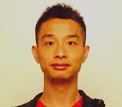
Title: Uncovering Cellular Dynamics of Mammalian Cardiac Regeneration Through Intravital Kilohertz Imaging of the Beating Heart
During development, the mammalian heart loses its potential to functionally regenerate after injury, resulting in heart disease being the number one cause of death across the globe. Intravital imaging of the beating heart is very challenging due to its anatomical location and rhythmic contraction. Current studies of mouse heart regeneration are largely limited to postmortem analysis of heart tissue to explore cellular activity and molecular mechanisms. In this proposal, we aim to combine a novel imaging window system designed and surgically implanted on the mouse chest by the Huang Lab at UCSF with the free-space angular-chirp-enhance delay (FACED) kilohertz-frame-rate microscopy system developed by the Ji Lab at UC Berkeley. With ultrafast fluorescent imaging of the beating heart in vivo, we aim to observe the dynamic behavior of single cardiomyocytes during the regenerative process after injury as well as track the formation of new vascular networks within the heart tissue that arise to compensate for ischemic injury. Long-term and high-resolution imaging of the mouse heart in vivo would allow the characterization of the behaviors and interactions of various cardiac cell types in their native environment during the regenerative process, uncovering unprecedented insights into the dynamic process of endogenous cardiac repair.
2022/23 Recipient
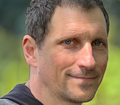
Title: Non-Invasive Risk Assessment of Dural Arteriovenous Fistulas using Displacement Spectrum (DiSpect) MRI
Dural arteriovenous fistulas (DAVFs) is a dangerous acquired vascular condition in the brain where several arteries with high blood pressure directly connect to the typically low-pressure veins and can cause life-threatening intracranial hemorrhage. DAVFs account for 10-15% of intracranial vascular malformations, commonly a result of head trauma, that are notoriously challenging to diagnose and impossible to risk stratify with conventional noninvasive imaging. X-Ray based Digital subtraction angiography (DSA) remains the gold standard for diagnosis and the only means for grading risk. However, DSA is an invasive procedure that has risks including life-threatening stroke. When DAVFs are present, pressures in the draining veins of the head elevate and can result in flow reversal in the cortical veins – the signature of a particularly high-risk condition. In recent years, there has been much progress in developing non-invasive MRI methods for determining vasculature structure, flow and perfusion. Despite these recent advances, noninvasive imaging is ill-suited for determining drainage patterns which, when dysfunctional, can have a profound clinical impact.
In this project, Professor Lustig and PhD candidate Ekin Karasan from UC Berkeley together with Professors Matthew Amans and David Saloner from UC San Francisco will develop non-invasive MRI methods for evaluating the risk of DAVFs. We have been developing uniquely suited MRI techniques that have huge potential for untangling the complex drainage of DAVFs. The aim of this proposal is clinical translation and application of these novel techniques to patients with DAVFs to identify not only the connections between the major draining veins and their tributaries, but also the presence of normal draining flow and high-risk reverse flow. Establishing the prevailing flow conditions will provide a non-invasive, decision-making tool for the neurointerventionalist that can guide if and when treatment is needed.
2021/22 Recipients
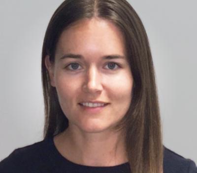
Title: Computational methods for causal genetic variant discovery in polygenic disease, with application to kidney disease
Genome-wide genetic variation affects our risks for complex polygenic diseases, but identifying the variants causal for disease risk and determining their mechanisms of action is an unsolved challenge in human genetics. Genome-wide association studies (GWAS) identify variants associated with disease, but most of these associations are non-causal. Since these associations predominantly fall in non-protein-coding regions of the genome, annotation of noncoding regulatory elements greatly improves fine-mapping of causal variants at associated loci. Deep learning methods trained on experimentally defined genome-wide epigenetic features can predict the effects of genetic variants on such regulatory elements, but optimizing these methods for use in GWAS fine-mapping and causal variant discovery remains a challenge.
In this project, the Ioannidis lab will collaborate with Profs. Jeremy Reiter and Gabriel Loeb at UCSF, combining computational and experimental expertise to address this challenge and advance methods for causal variant discovery, with application to kidney disease. We are using single-cell experimental measurements of chromatin accessibility, histone modifications, and gene expression in primary kidney cells to train deep learning models to predict the cell-type specific effects of genetic variants on these molecular phenotypes. We will advance the design and training of these models to improve their sensitivity to changes caused by single nucleotide variants, as well as their performance in disease-relevant regions of the genome containing kidney-specific regulatory elements. The methods developed here will not only advance our understanding of the genetic mechanisms underlying kidney disease, but will be widely applicable across many complex polygenic diseases.
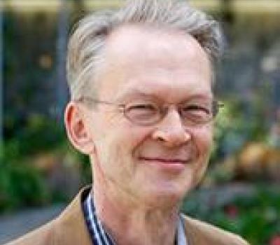
Title: Quantifying Invasive Tumor Volume by Deep Learning to Improve Malignant Melanoma Prognosis
Melanoma is the deadliest skin cancer with the fastest rising incidence of cancer in the United States. The most important predictor of melanoma patient survival is the volume of invasive tumor at the first biopsy. However, the current standard of care for outcome prediction is to manually measure a single-dimension tumor thickness, which acts as a proxy for volume. The predictive accuracy of this single measurement depends on a host of subjectively defined measurements which can lead to incorrect risk assessment and management planning. Recent advances in deep learning as applied to digital pathology will allow us to objectively assess the entirety of the invasive tumor and its interrelated variables. Here, we propose to use deep learning methods to automatically and accurately quantify the invasive tumor’s cross-sectional area, at the sensitivity of single tumor cells. We will calculate the relationship between the automatically calculated invasive melanoma areas to patient survival, and develop a survival prediction algorithm. We hypothesize that this efficient and reproducible method will better predict patient survival than current methods.
2020/21 Recipient
Title: Wound closing and healing devices: Design, computation, and experimental validation
Injuries to the human body are a common occurrence, from accidental paper cuts to complex surgeries. These cutaneous and sub-cutaneous wounds oftentimes require wound care to facilitate the healing of the injured tissue, especially in chronic injuries and surgical procedures. Current methods and materials designed for wound closure present an invasive approach to seal wounds and often require skilled practitioners for a successful application. Invasive methods and incorrect application of these materials can lead to delayed healing, cosmetic deformities upon healing, and lead to prolonged inflammation.
In this research, Gu Lab from UC Berkeley together with Desai Lab from UCSF will look to design, optimize, and fabricate novel non-invasive wound closing materials through a combined computational and experimental approach for stimulated wound closure using temperature and humidity. The proposed approach will use materials adhered to the surface of the skin that will change shape when stimulated and automatically close the wound during the process. The combined expertise in biomaterials, computational mechanics, and additive manufacturing between the two labs is well-suited to tackle the challenges of developing new materials for medical applications. Understanding these material design parameters for wound sealing and healing can guide the design of stimuli-responsive materials for other uses, such as self-actuating components for robotics and personalized, self-healing textiles and body armor applications.
2019/20 Recipient
Title: Clinical and Economic Endpoints for Assessing Employer Strategies to Shift Patients from Hospital-based to Freestanding Ambulatory Surgery
This project will support a larger multi-year research study of clinical and economic performance for major surgical, medical, and diagnostic procedures in freestanding (ASC) and hospital-based (HOPD) settings. We will identify the procedures to be compared across ASCs and HOPDs, and of particular importance, the relevant clinical endpoints. We will also develop economic endpoints, such as total payment and patient cost sharing; and utilization endpoints, such as discharge disposition, length of stay, emergency department visits, and readmissions.
This proposed project will combine the economic expertise of the Berkeley faculty and the clinical knowledge and expertise of Professor Sanket Dhruva from the UCSF School of Medicine, along with graduate student research assistants on both campuses. We are hopeful that this collaboration will establish the pilot results that can be used for a successful multi-year collaboration between the two campuses.
2018/19 Recipient
Title: Super-resolution microscopy for living cells
As one of the newest breakthroughs in light microscopy, the invention of super-resolution microscopy brings the spatial resolution towards the size of a protein molecule (~ 10 nm), thus generating enormous excitement in biologists. Among super-resolution microscopy techniques, the single-molecule-switching-based approach, commonly known as STORM or PALM, achieves super-resolution by activating and sampling small fractions of fluorophores in each camera frame. Despite the achievements in 3D and multicolor super-resolution imaging, the development of live cell STORM/PALM has been hindered by its requirement for a large number of camera frames and high excitation light intensity, the duration of observation becomes one of the final obstacles for it to be broadly applicable to the study of subcellular dynamic processes. Here, combining the expertise of Dr. Bo Huang at UCSF in super-resolution microscopy technique development and the expertise of hosting Dr. Laura Waller at UC Berkeley in computational imaging, we propose a computational approaches to address this issue. We will create new algorithms that can reconstruct high quality super-resolution images from noisy snapshots in a continuous movie, as well as algorithms to infer the underlying cellular structures from incompletely sampled snapshots utilizing prior knowledge of the structures. These methods should increase the observation duration of live STORM/PALM by more than one order of magnitude. We are confident that our research will have far reaching impact given the sheer number of biological problems awaiting visualization tools.
2017/18 Recipients
Title: The structural basis of compensatory mutations using cryo-electron microscopy
Mutations in proteins can exhibit non-additivity (epistasis). For example, mutation X can be deleterious in one background, yet neutral or beneficial when combined with mutation Y. An extreme example of this non-additivity is the case of “compensated pathogenic deviations” where mutations that are causal of disease in humans are tolerated as the wild type residue in other species. Using bioinformatics methods and in vivo selections we have identified compensatory mutations in the metabolic enzyme glutamine synthetase. Our working hypothesis is the disease mutations bias the conformational ensemble of the enzyme towards a non-functional oligomeric filament and that the compensatory mutations restore the enzyme towards the non-filamentous state.
James Fraser is an assistant professor in the Department of Chemistry at UC Berkeley and a Faculty Scientist at the Lawrence Berkeley National Laboratory. His group uses insights from cellular biophysics, physical chemistry, and materials science to study key aspects of signal transduction processes in cell membranes.
Title: Real-time Chromatin Condensation by High-Speed Atomic Force Microscopy
The compaction of eukaryotic DNA into chromatin enables both the accommodation of a large amount DNA into a constrained nuclear space as well as regulation of DNA based transactions such as transcription and replication. Thus regions of the genome with highly compacted chromatin structures are silenced for expression, whereas those with relaxed open structures correspond to actively expressed genes. The basic repeating unit of chromatin is a nucleosome, which contains ~147 bp of DNA wrapped around an octamer of histone proteins. How chromatin adopts and retains specific compacted three-dimensional (3D) structures has received substantial attention for several decades. Yet, there is a lack of consensus about the factors that govern chromatin compaction and the underlying molecular mechanisms. The Bustamante and Narlikar laboratories, propose to use high speed atomic force microscopy (HS-AFM) to follow in real-time the transition of chromatin primary structures into compacted 3D ones. In preliminary experiments, nucleosomal arrays provided by Narlikar lab have been visualized at high spatial (1-2 nm) and temporal resolution (up to 100 msec per frame) in solution in the Bustamante lab. In this proposal we aim at expanding these studies by directly visualizing the effects of different ions and protein regulators on the dynamics of chromatin folding and on the resultant 3D chromatin structures. The simultaneous temporal and spatial resolution provided by the HS-AFM will yield unprecedented insight into the process by which chromatin attains its higher order organization.
2016/17 Recipients
Title: Operant conditioning of abnormal cortical activity in Parkinson’s disease
In collaboration with colleagues at UCSF, and with support from the Sackler Program, Jose Carmena will pilot a novel approach to exploring the importance of specific brain rhythms in producing motor deficits in Parkinson’s disease, by training subjects to change those rhythms in an operant conditioning paradigm and measuring associated changes in parkinsonian motor signs.
Jose M. Carmena is a professor of Electrical Engineering and Neuroscience at UC Berkeley, and Co-Director of the Center for Neural Engineering and Prostheses at UCB and UCSF. His expertise is in brain-machine interfaces, an area of high relevance for normal and disordered sensorimotor function. His lab is known for seminal studies showing how neural plasticity relates to the acquisition and consolidation of neuroprosthetic skills in the mammalian brain, and is also very active in the development of neurotechnology and in closed-loop decoding algorithms for neuroprosthetic control. Dr. Carmena received a number of honors, including the Bakar Fellow award, the IEEE-EMBS Early Career Achievement Award, the Aspen Brain Forum Prize in Neurotechnology, the NSF CAREER Award, the Alfred P. Sloan Fellowship, and the Hellman Family Faculty Fund Award.
Title: Organs on a chip – the future of personalize medicine
Kevin Healy is an innovator working at the interface between stem cells and materials science to develop dynamic engineered systems to explore both fundamental biological phenomena and new applications in translational medicine. His group currently conducts research on human tissue models for drug discovery (e.g. organs-on-chips), which is traditionally hampered by high failure rates attributed to reliance on non-human animal models that poorly replicate human disease states. In collaboration with colleagues at UCSF, Healy’s group is developing multi-organ systems to circumvent this problem by understanding the fundamental relations between critical organs (i.e. heart, liver) as a complex living system, and to accurately predict the tissue- and system-specific effect of drugs before human administration. The Sackler sabbatical exchange will foster new lines of inquiry and accelerate the research program so that both healthy and disease-specific multi-organ systems become indispensable for drug discovery and patient specific medicine.
Dr. Healy is the Jan Fandrianto and Selfia Halim Distinguished Professor in Engineering at the University of California, Berkeley in the Departments of Bioengineering, and Materials Science and Engineering.
Title: Neuroeconomic Assessment of Decision-Making: Translational and Commercial Applications
The Sackler award will leverage the unparalleled strength of UCB and UCSF in basic and clinical neurosciences, as well as the deep knowledge of fostering entrepreneurship excellence at Berkeley-Haas so as to (1) bring recent scientific advances in understanding neural basis of financial and social decision-making to clinical settings, and to (2) explore potential commercial
applications of this knowledge.
Ming Hsu’s lab takes an interdisciplinary approach to study the biological basis of economic and consumer decision-making at a number of different levels of analyses, including: (i) brain regions mediating computations involved in different aspects of decision-making, (ii) the underlying molecular, cellular, and genetic mechanisms, and (iii) how real-world outcomes are associated with variations in these processes. Dr. Hsu is Associate Professor at the Haas School of Business and Helen Wills Neuroscience Institute, UC Berkeley. He is the recipient of awards from the NIH, Hellman Family Family Fund, Robert Wood Johnson Foundation, and Risk Management Institute.
2015/16 Recipients
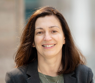
Title: Understanding Substrate Selectivity and Function of RNA Methylation
In collaboration with colleagues at UC Berkeley, and with support from the Sackler Program, Danica Fujimori will investigate physiological relevance of RNA methylation and will develop cell engineering applications that utilize modified RNAs.
Dr. Fujimori is an associate professor in the Department of Cellular and Molecular Pharmacology, and the Department of Pharmaceutical Chemistry at UC San Francisco. Her research aims to develop a better understanding of the mechanisms, regulation, and biological functions of methyl group addition and removal in proteins and RNA. The enzymatic regulation of methyl group modifications provides an opportunity for therapeutic intervention for a wide range of diseases.
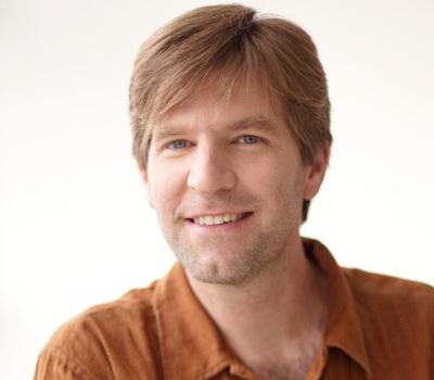
Title: Mechanical Regulation of RTKs in Cancer
A combination of discoveries by the Groves Lab and their colleagues at UCSF have led to new insights into how cancer cells acquire resistance to the drugs used to destroy them. Unfortunately many cancer patients see their cancer at first respond positively to drug treatment, only to return later with a newfound resistance to the cancer drugs. Support from the Sackler Program will allow this team to develop new ideas and acquire critical first proof-of-principle experimental data into how cancer cells develop this resistance.
Jay Groves is a professor in the Department of Chemistry at UC Berkeley and a Faculty Scientist at the Lawrence Berkeley National Laboratory. His group uses insights from cellular biophysics, physical chemistry, and materials science to study key aspects of signal transduction processes in cell membranes.
2014/15 Recipients

Title: Recognizing the Enemy Within: Mechanisms that License Genome Defense
Hiten Madhani is a professor of biochemistry and biophysics at UCSF. Dr. Madhani’s group investigates the biology of the budding yeast Cryptococcus neoformans. Infection by this opportunistic yeast pathogen is most common cause of fungal meningitis worldwide, and is responsible for ~1/3 of deaths in HIV/AIDS. His laboratory is interested in how Cryptococcus survives in the mammalian host, how it evades the host immune system, and how it responds to small molecules. The Madhani group also uses Cryptococcus to investigate basic mechanisms of gene regulation with a focus on chromatin and small RNA-based control of selfish genetic elements.
Dr. Madhani holds BS and MS degrees from Stanford University and MD and PhD degrees from UCSF. His received postdoctoral training at Whitehead Institute/MIT. He has received research awards from the David and Lucille Packard Foundation, the Burroughs-Welcome Fund, Leukemia and Lymphoma Society and the National Institutes of Health. In 2014 he was elected to the American Academy of Microbiology.
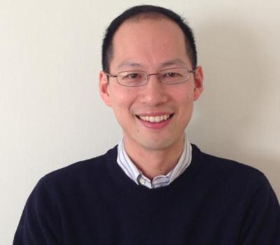
Title: Recombinant Antibodies to Target Copper Homeostasis as an Anticancer Therapeutic Strategy
Research in the Chang lab is focused on chemical biology and inorganic chemistry, with particular interests in molecular imaging and catalysis applied to neuroscience, metabolic diseases, and sustainable energy.
Dr. Chang's work has been honored by awards from the Dreyfus, Beckman, Sloan, and Packard Foundations, Amgen, Astra Zeneca, and Novartis, Technology Review, ACS (Cope Scholar, Eli Lilly, Nobel Laureate Signature, Baekeland), RSC (Transition Metal Chemistry), and SBIC.
2013/14 Recipients
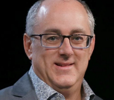
Scott Baraban is a professor of neurological surgery and William K. Bowes Jr. Endowed Chair in Neuroscience Research at the University of California, San Francisco. Dr. Baraban’s lab studies the cellular and molecular basis of epilepsy, a devastating neurological disorder afflicting nearly 3 million Americans. While some seizures can be controlled with available medications, a large number of epilepsy patients are medically intractable. Combining pharmacology, genetics, electrophysiology and unique zebrafish models of intractable epilepsy they are identifying new treatments for these patients.
Dr. Baraban is the recipient of awards from the Esther and Joseph Klingenstein Fund, the Sandler Family Supporting Foundation, the UCSF Innovation in Basic Science Award, as well as a EUREKA grant from the NIH.
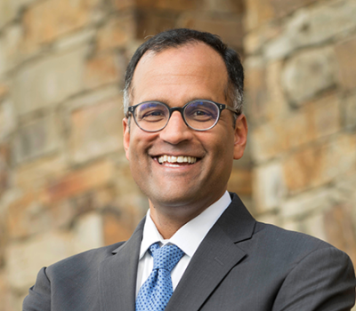
Sanjay Kumar is a professor of bioengineering at UC Berkeley. Dr. Kumar’s research team is working to understand how biophysical inputs control cell and tissue behavior and exploiting this knowledge to engineer biological function. This is applied to stem cell and cancer biology/therapeutics, primarily in the context of the central nervous system.
Dr. Kumar has received a number of honors, including the Presidential Early Career Award for Scientists and Engineers (PECASE), the NIH Director’s New Innovator Award, the Arnold and Mabel Beckman Young Investigator Award, the NSF CAREER Award, and the Hellman Family Faculty Fund Award.
