Innovative Design Achieves Tenfold Better Resolution for Functional MRI Brain Imaging
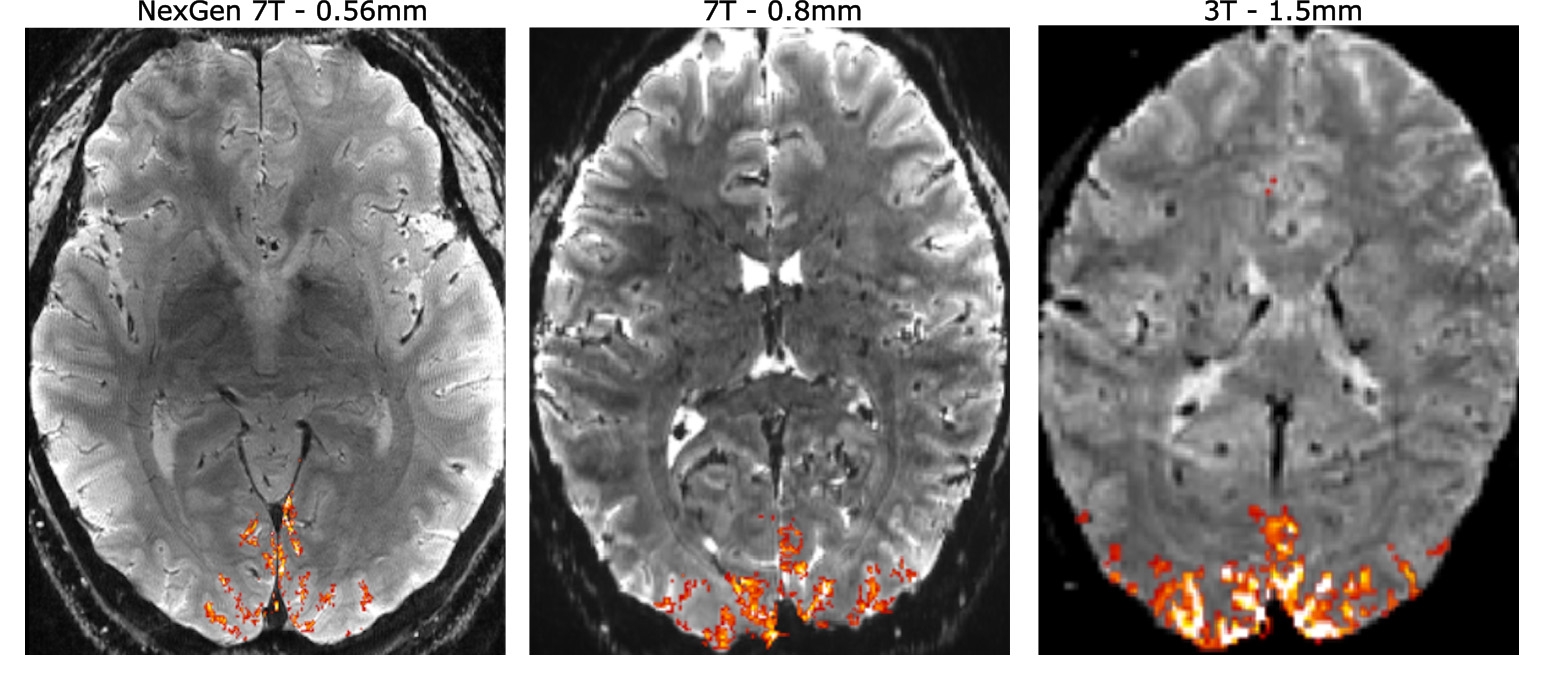
An intense international effort to improve the resolution of magnetic resonance imaging (MRI) for studying the human brain has culminated in an ultra-high resolution 7 Tesla scanner that records up to 10 times more detail than current 7T scanners and over 50 times more detail than current 3T scanners, the mainstay of most hospitals.
The dramatically improved resolution means that scientists can see functional MRI (fMRI) features 0.4 millimeters across, compared to the 2 or 3 millimeters typical of today's standard 3T fMRIs.
"The NexGen 7T scanner is a new tool that allows us to look at the brain circuitry underlying different diseases of the brain with higher spatial resolution in fMRI, diffusion and structural imaging, and therefore to perform human neuroscience research at higher granularity. This puts UC Berkeley at the forefront of human neuroimaging research," said David Feinberg, the director of the project to build the scanner, acting professor at the Helen Wills Neuroscience Institute at the University of California, Berkeley, and president of Advanced MRI Technologies (AMRIT), a research company based in Sebastopol, California.
"The ultra-high resolution scanner will allow research on underlying changes in brain circuitry in a multitude of brain disorders, including degenerative diseases, schizophrenia and developmental disorders, including autism spectrum disorder."
This next generation or NexGen 7T MRI scanner is described in a paper that will be published Nov. 27 in the journal Nature Methods.
The improved resolution will help neuroscientists probe the neuronal circuits in different regions of the brain's neocortex and allow researchers to track signals propagating from one area of the cortex to another as we think and reason, and perhaps discover underlying causes of developmental disorders. This could lead to better ways of diagnosing brain disorders, perhaps by identifying new biomarkers that would allow diagnosis of mental disorders earlier or, more specifically, in order to choose the best therapy.
"Normally, MRI is not fast enough at all to see the direction of the information being passed from one area of the brain to another," Feinberg said. "The scanner’s higher spatial resolution can identify activity at different depths in the brain’s cortex to indirectly reveal brain circuitry by differentiating activity in different cell layers of the cortex."
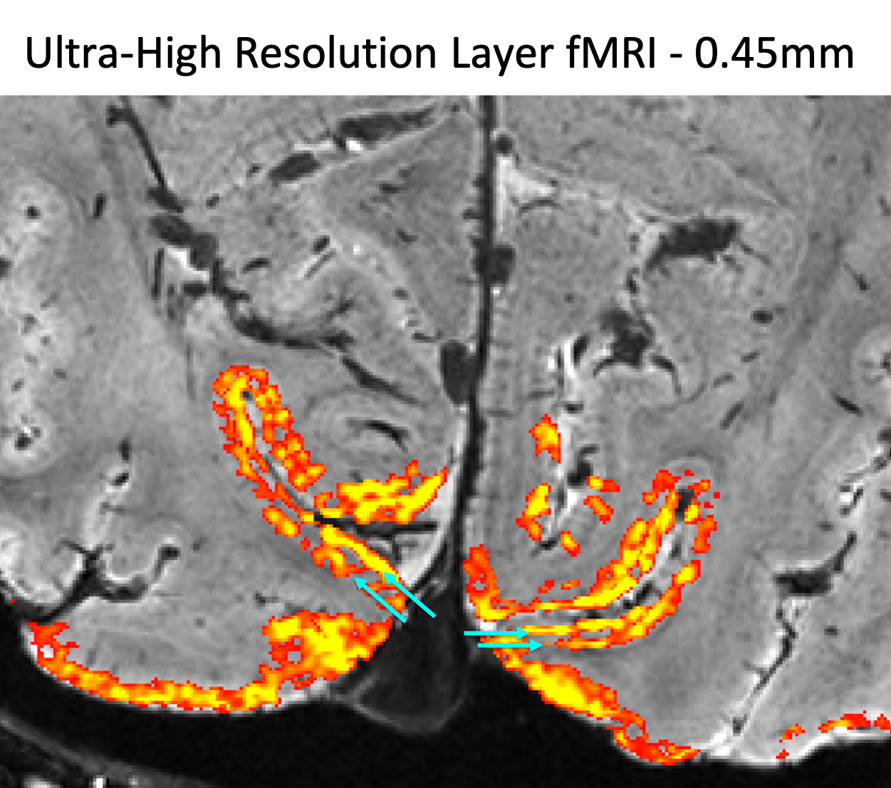
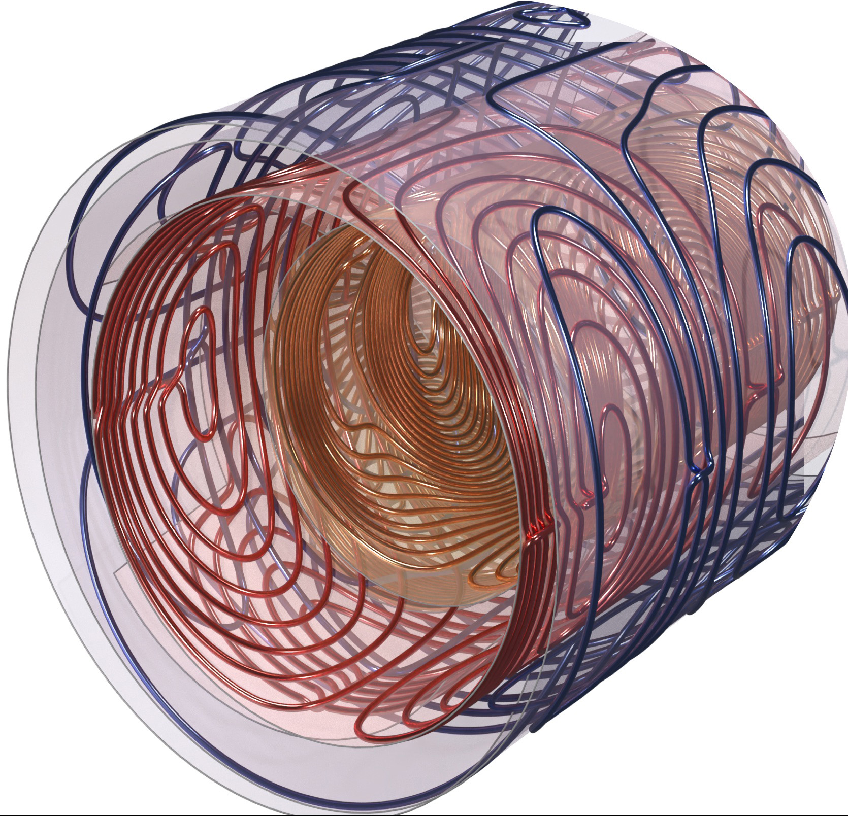
This is possible because neuroscientists have found in vision brain areas that the superficial and deepest cortex layers (blue arrows in image on right) incorporate "top-down" circuits, that is, they receive information from higher cortical brain areas, whereas the middle cortex involves "bottom-up" circuitry, receiving input to the brain from our senses. Pinpointing the fMRI activity to a specific depth in the cortex lets neuroscientists track the flow of information throughout the brain and cortex.
With the higher spatial resolution, neuroscientists will be able to home in on the activity of something on the order of 850 individual neurons within a single voxel — a 3D pixel — instead of the 600,000 recorded with standard hospital MRIs, said Silvia Bunge, a UC Berkeley professor of psychology who is one of the first to use the NexGen 7T to conduct research on a human brain.
"We were able to look at the layer profile of the prefrontal cortex, and it's beautiful," said Bunge, who studies abstract reasoning. "It's so exciting to have this state-of-the-art, world-class machine."
For William Jagust, a UC Berkeley professor of public health who studies the brain changes associated with Alzheimer's disease, the improved resolution could finally help connect the dots between observed changes due to Alzheimer’s that occur in the brain — abnormal clumps of protein called beta amyloid and tau — and changes in memory.
"We know that part of the memory system in the brain degenerates as we get older, but we know little about the actual changes to the memory system — we can only go so far because of the resolution of our current MRI systems," said Jagust. "With this new scanner, we think we're going to be able to take apart a lot more carefully exactly where things have gone wrong. This could help with diagnosis or predicting outcomes in normal people."
Jack Gallant, a UC Berkeley professor of psychology, hopes the scanner will help neuroscientists discover how functional changes in the brain lead to developmental and mental disorders such as dyslexia, autism and schizophrenia, or that result from neurological disorders, such as dementia and stroke.
"Mental disorders have an enormous impact on individuals, families and society. Together they represent about 10% of the U.S. GDP. Mental disorders are fundamentally disorders of brain function, but functional measures are not used currently to diagnose most brain disorders or to look to see if a treatment's working. Instead, these disorders are diagnosed behaviorally. This is a weak approach, because there are a lot of different mental brain states that can lead to exactly the same behavior," Gallant said. "What we need is more powerful MRI machines like this so that we can map, at high resolution, how information is represented in the brain. To me this is the big potential clinical benefit of ultra-high resolution MRI."
BRAIN Initiative
The breakthrough came about through an initial $13.4 million in funding from the Brain Research through Advancing Innovative Neurotechnologies (BRAIN) Initiative of the U.S. National Institutes of Health (NIH). The initiative aims to develop new technologies that will produce a dynamic picture of the brain showing how individual cells and complex neural circuits interact across the brain and over time.
Additional funding from UC Berkeley's Chancellor's Office and the Weill Neurohub brought the total funding to over $22 million, which allowed Feinberg to assemble a multidisciplinary team of academics and leading scientists at the multinational corporation Siemens Healtheneers, a major manufacturer of hospital and research MRI scanners; MR CoilTech Limited of Glasgow, Scotland, maker of transmitter and receiver detector coils used in MRI to generate and record signals; and AMRIT, a designer of imaging pulse sequences that control the scanner hardware.
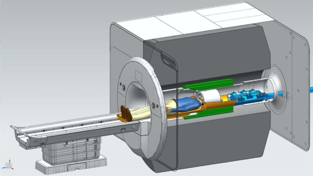
Incorporating newly developed hardware technology from those groups, Siemens collaborated with Feinberg’s team to rebuild a conventional 7 Tesla MRI scanner delivered to UC Berkeley in 2000 to improve the spatial resolution in pictures captured during brain scans.
"There's been a large increase throughout the world of sites that use 7T MRI scanners, but they were mostly for development and were difficult to use," said Nicolas Boulant, a physicist visiting from the NeuroSpin project at the University of Paris in Saclay, where he leads the team that operates the world's only 11.7 Tesla MRI scanner, the strongest magnetic field employed to date. "David's team really put together many ingredients to make a quantum leap at 7 Tesla, to go beyond what was achievable before and gain performance."
Boulant hopes to adapt some of the new ingredients in the NexGen 7T — in particular, redesigned gradient coils — to eventually achieve even better resolution with the 11.7 Tesla MRI scanner. The gradient coils generate a rising magnetic field across the brain so that each part of the brain sees a different field strength, which helps to precisely map brain activity.
"The higher the magnetic field, the more difficult it is to really grab the potential promised by these higher-field MRI scanners to see finer details in the human brain," he said. "You need all this peripheral equipment, which needs to be on steroids to meet those promises. The NexGen 7T is really a game-changer when you want to do neuro MRI."
To reach higher spatial resolution, the NexGen 7T scanner had to be designed with a greatly improved gradient coil and with larger receiver array coils — which detect the brain signals — using from 64 to 128 channels to achieve a higher signal-to-noise ratio (SNR) in the cortex and faster data acquisition. All these improvements were combined with a higher signal from the ultra-high field 7T magnet to achieve cumulative gains in the scanner performance.
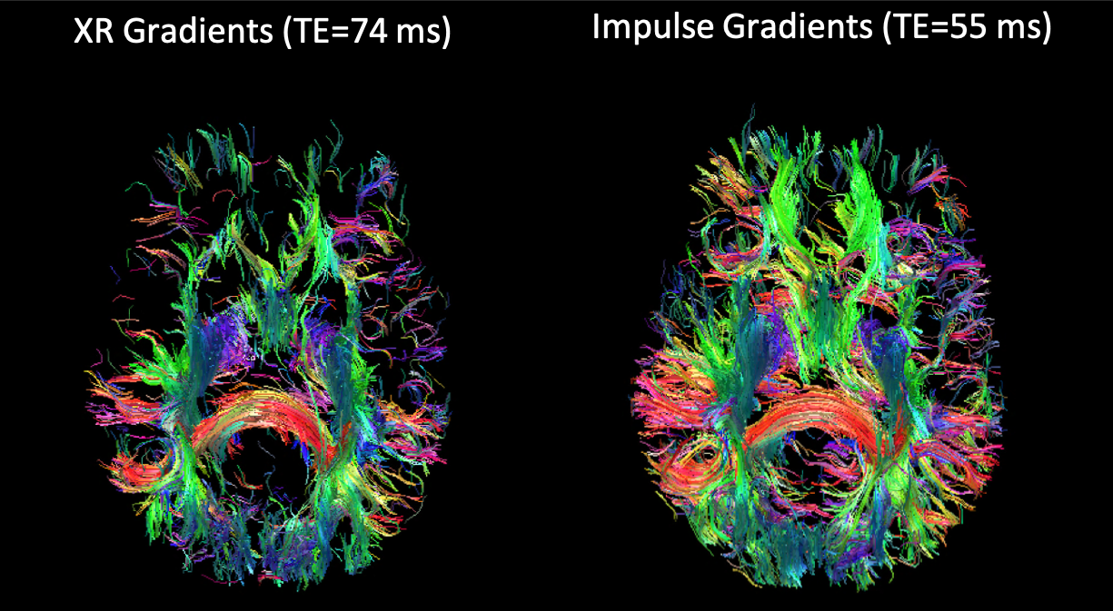
The extremely powerful gradient coil is the first to be made with three layers of wire windings. Designed by Peter Dietz at Siemens in Erlangen, Germany, the "Impulse" gradient has 10 times the performance of gradient systems in current 7T scanners. Mathias Davids, then a physics graduate student at Heidelberg University in Mannheim, Germany, and a member of Feinberg’s team, collaborated with Dietz in performing physiologic modeling to allow a faster gradient slew rate — a measure of how quickly the magnetic field changes across the brain — while remaining under the neuronal stimulation thresholds of the human body.
"It's designed so that the gradient pulses can be turned on and off much quicker — in microseconds — to record the signals much quicker, and also so the much higher amplitude gradients can be utilized without stimulating the peripheral nerves in the body or stimulating the heart, which are physiologic limitations," Feinberg said.
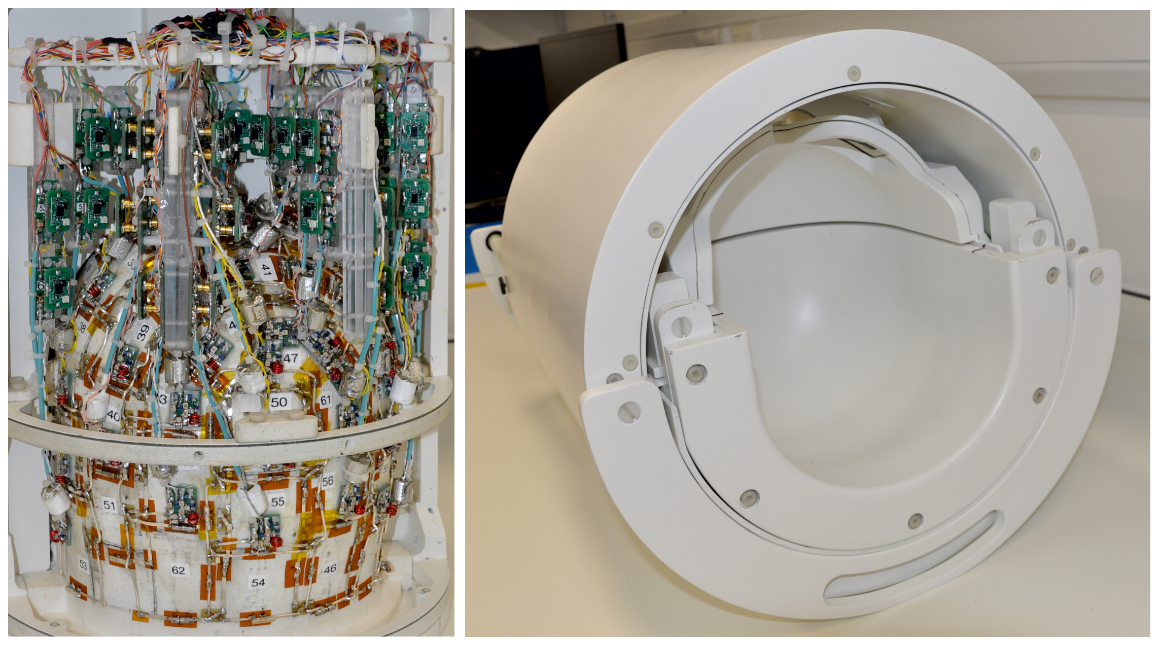
A second key development in the scanner, Feinberg said, is the 128-channel receiver system that replaces the standard 32 channels. The large receiver coil arrays built by Shajan Gunamony of MR CoilTech in Glasgow, UK, gave a higher signal-to-noise ratio in the cerebral cortex and also provided higher parallel imaging acceleration for faster data acquisition to encode large image matrices for ultra high resolution fMRI and structural MRI.
To take advantage of the new hardware technology, Suhyung Park, Rüdiger Stirnberg, Renzo Huber, Xiaozhi Cao and Feinberg designed new pulse sequences of precisely timed gradient pulses to rapidly achieve ultra high resolution. The smaller voxels, measured in units of cubic millimeters and less than 0.1 microliter, provide a 3D image resolution that is 10 times higher than that of previous 7T fMRIs and 125 times higher than the typical hospital 3T MRI scanners used for medical diagnosis.
Voxels matter
The most common MRI scanners employ superconducting magnets that produce a steady magnetic field of 3 Tesla — 90,000 times stronger than Earth's magnetic field.
“A 3T scanner can resolve fMRI detail only about 2 mm to 3 mm across, whereas the entire thickness of the six cortical layers composing the brain gray matter is only 1.5 to 4.5 mm thick. That makes it impossible to see and study the function of microcircuits — which are only 0.5 mm across — that are responsible for specific actions,” Gallant said.
In contrast, fMRI focuses on blood flow in arteries and veins and can vividly distinguish oxygenated hemoglobin funneling into working areas of the brain from deoxygenated hemoglobin in less active areas. This allows neuroscientists to determine which areas of the brain are engaged during a specific task.
But again, the 3 mm resolution of a 3T fMRI can distinguish only large veins, not the small ones that could indicate activity within microcircuits.
The NexGen 7T will allow neuroscientists to pinpoint activity within the thin cortical layers in the gray matter, as well as within the narrow column circuits that are organized perpendicular to the layers. These columns are of special interest to Gallant, who studies how the world we see is represented in the visual cortex. He has actually been able to reconstruct what a person is seeing based solely on recordings from the brain's visual cortex.
"The machine that David has built, in theory, should get down to 500 microns, or something like that, which is way better than anything else — we're very near the scale you would want if you're getting signals from a single column, for example," Gallant said. "It's fantastic. The whole thing about MRI is how big is the little volumetric unit, the voxel, the three-dimensional pixel that you're recording from. That's the only thing that matters."
For the moment, NexGen 7T brain scanners must be custom-built from regular 7T scanners. The cost should be considerably lower than the $22 million required to build the first one, however. These funds came not only from the BRAIN Initiative, but also from UC Berkeley funds through the Helen Wills Neuroscience Center, with which Feinberg, Bunge, Gallant and Jagust are affiliated.
Feinberg said that UC Berkeley’s NexGen 7T scanner technology will be disseminated by Siemens and MR CoilTech Ltd.
"My view is that we may never be able to understand the human brain on the cellular synaptic circuitry level, where there are more connections than there are stars in the universe," Feinberg said. " But we are now able to see signal patterns of brain circuits and begin to tease apart feedback and feed forward circuitry in different depths of the cerebral cortex. And in that sense, we will soon be able to understand the human brain organization better, which will give us a new view into disease processes and ultimately allow us to test new therapies. We are seeking a better understanding and view of brain function that we can reliably test and reproducibly use noninvasively."
Other co-authors of the paper are Alexander Beckett of Advanced MRI Technologies; Chunlei Liu of UC Berkeley's Helen Wills Neuroscience Institute; An (Joseph) Vu of UC San Francisco; Lawrence Wald, Bernhard Gruber, Jon Polimeni and Jason Stockmann of the A. A. Martinos Center for Biomedical Imaging at Massachusetts General Hospital; Kawin Setsompop of Stanford University in California; Rudiger Sternberg of the German Center for Neurodegenerative Diseases in Bonn, Germany; Laurentius (Renzo) Huber of Maastricht University in the Netherlands; and Suhyung Park at Chonnam National University, South Korea.
The work was supported by BRAIN Initiative grants through the NIH (U01-EB025162, R01-322 MH111444) and other NIH grants (P41-EB030006, NIH R44-MH129278), as well as by funds from UC Berkeley's Chancellor's Office and the Weill Neurohub.