
Research Bio
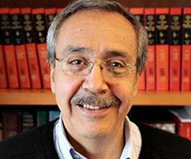
Carlos Bustamante is a Professor in the Department of Physics, the Department of Chemistry, and the Department of Molecular and Cell Biology. He is the Raymond and Beverly Sackler Professor in Biophysics.
Research in the Bustamante Lab is focused on the structural characterization of nucleo-protein assemblies. The structure of chromatin and the global structure of protein-nucleic acid complexes relevant to the molecular mechanisms of control of transcription in prokaryotes are investigated using high resolution scanning force microscopy (SFM). This microscope, also known as Atomic Force Microscope (AFM) works by scanning a tip over the sample to sense the topography of the surface, thus functioning in much the same way than old record players. In addition, they are studying the elastic response of long linear polymers, the forces responsible for maintaining the tertiary structure of proteins, and the mechanical properties of molecular motors, using methods of single molecule manipulation such as laser tweezers and the SFM.
Current Projects
The laboratory is involved in the study of the structural basis of protein-DNA interactions and their relevance in the processes of control of gene expression. In prokaryotes, and especially in eukaryotes, replication and transcription regulation involve the interaction of many specialized protein factors at regulator locations on the sequence to ensure correct sequence recognition, initiation, processivity, fidelity, and kinetic control. They wish to understand the multiple structural, spatial, and functional relationships among these regulatory factors. They are using the SFM as a high resolution tool to image initiation and elongation transcription complexes of E. coli RNA polymerase to characterize the spatial relationships between the enzyme and the DNA template. They are also beginning to investigate what structural changes are negotiated between RNA polymerase and chromatin during transcription. To this end, they are using the SFM to image complexes of nucleosome-containing DNA fragments carrying a promoter and a terminator upstream and downstream of the nucleosome positioning sequence, respectively. They plan to compare the behavior of various prokaryotic and eukaryotic polymerases as they transcribe through the nucleosome, to investigate whether transcription through a nucleosome is an inherent property of the core particle, or a property of each enzyme itself, and to characterize various intermediates of the translocation process.
Our laboratory is also working actively in the development of methods of single-molecule manipulation, including the use of SFM cantilevers, optical or laser tweezers, and magnetic beads to investigate the mechanical properties of macromolecules. In one project, they first tether a single protein molecule of T-4 lysozyme between a surface and the end of an SFM cantilever. They can then separate the surfaces in a controlled fashion to induce the mechanical unfolding of the molecule to characterize the nature, range, and strength of the forces that maintain its three-dimensional structure. Our objective is to carry out the unfolding of the molecule at equilibrium so as to obtain the potential energy function of the molecule as a function of the mechanical extension. This function represents the most complete description of the folded state of the protein. They plan to investigate how external conditions in the medium, i.e. temperature, denaturant concentration, etc., or point-directed mutations affect the shape of the potential energy function.
Finally, our laboratory is also engaged in the study of DNA-binding molecular motors (RNA polymerase, DNA polymerase, etc.) using optical tweezers to investigate the dynamics of these molecules during translocation, as well as the effect of external force load and nucleotide tri-phosphate concentration on their power and force generation. In parallel, they are developing both microscopic (chemical ratchet-type) and phenomenological models of molecular motors which will be tested experimentally. They believe that single molecule experiments can provide a unique look into the molecular mechanisms responsible for the mechano-chemical conversion process in these protein machines.
Research Expertise and Interest
nanoscience, structural characterization of nucleo-protein assemblies, single molecule fluorescence microscopy, DNA-binding molecular motors, the scanning force microscope, prokaryotes
In the News
Can Synthetic Polymers Replace the Body’s Natural Proteins?
Bustamante awarded Biophysical Society Honors
Researchers Find that Viral Packaging Motor Rotates DNA and Adapts to Changing Conditions
While a virus, essentially, may be nothing more than a dollop of DNA packed into a protective coating of protein called a capsid, the packaging of that DNA is critical. The molecular motors that drive this DNA packaging process, however, have remained almost as enigmatic as the viruses themselves.
UC Berkeley, Berkeley Lab announce Kavli Energy NanoScience Institute
The Kavli Foundation has endowed a new institute at the University of California, Berkeley, and the Lawrence Berkeley National Laboratory (Berkeley Lab) to explore the basic science of how to capture and channel energy on the molecular or nanoscale and use this information to discover new ways of generating energy for human use.
Carlos Bustamante honored with Vilcek Prize
Carlos Bustamante, a professor of molecular and cell biology and of physics and chemistry, has been awarded the 2012 Vilcek Prize

